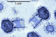
Mimiviridae is a family of viruses. Amoeba and other protists serve as natural hosts. The family is divided in up to 4 subfamilies.[1][2][3][4] Viruses in this family belong to the nucleocytoplasmic large DNA virus clade (NCLDV), also referred to as giant viruses.
| Mimiviridae | |
|---|---|
 | |
| Tupanvirus | |
| Virus classification | |
| (unranked): | Virus |
| Realm: | Varidnaviria |
| Kingdom: | Bamfordvirae |
| Phylum: | Nucleocytoviricota |
| Class: | Megaviricetes |
| Order: | Imitervirales |
| Family: | Mimiviridae |
| Subfamilies and genera | |
| |
Mimiviridae is the sole recognized member of order Imitervirales. Phycodnaviridae and Pandoraviridae of Algavirales are sister groups of Mimiviridae in many phylogenetic analyses.[5]
History
The first member of this family, Mimivirus, was discovered in 2003,[6] and the first complete genome sequence was published in 2004.[7] However, the mimivirus Cafeteria roenbergensis virus[8] was isolated and partially characterized in 1995,[9] although the host was misidentified at the time, and the virus was designated BV-PW1.[8]
Taxonomy
Group: dsDNA
- Family: Mimiviridae
Family Mimiviridae is currently divided into three subfamilies.[2][3][10]
- One subfamily (genus Mimivirus, proposed names: Megavirinae or Megamimivirinae) is divided into three "lineages":
- A — Mimivirus group: includes Acanthamoeba polyphaga Mimivirus, Hirudovirus, Mamavirus, Kroon virus, Lentille virus, Terra2, Niemeyer virus, Samba virus.[11][12]
- B — Moumouvirus group: includes Moumouvirus, Saudi moumouvirus, Moumouvirus goulette, Monve virus (aka Moumouvirus monve), and Ochan virus.[13][11][14][12]
- C — Courdo11 virus group: includes Mont1,[11] Courdo7, Courdo11, Megavirus chilensis, LBA111, Powai lake megavirus and Terra1.[15][16]
- The majority of Mimiviridae appear to belong to this subfamily (Mimiviruses).[10]
- It is sometimes also referred to as Mimiviridae group I.[17]
- The second subfamily (Cafeteriavirus or Mimiviridae group II) includes the Cafeteria roenbergensis virus (CroV).[8]
- The Klosneuvirinae have been proposed as a third subfamily and are divided into four "lineages": Klosneuvirus, Indivirus, Catovirus and Hokovirus.[3] They seem to be closely related to the Mimivirus subfamily rather than the Cafeteriavirus subfamily (and so might be summarized in Mimivirus group I as well).[3] The first isolate from this group is Bodo saltans virus infecting the kinetoplastid Bodo saltans.[18]
- Tupanvirus strains have been discussed to comprise a sister group of mimiviruses.[4]
Furthermore, it has been proposed either to extend Mimiviridae by an additional tentative group III (subfamily Mesomimivirinae) or to classify this group as a sister family Mesomimiviridae instead,[19] comprising legacy OLPG (Organic Lake Phycodna Group). This extension (or sister family) may consist of the following:
- Phaeocystis globosa virus (PgV, represented by PgV-16T strain) and Phaeocystis pouchetii virus (PpV, e. g. PpV 01)
- "Organic Lake Phycodnavirus" 1 and 2 (OLV1, OLV2, hosts of Organic Lake virophage)
- "Yellowstone Lake Mimivirus"[12][20] aka "Yellowstone Lake Phycodnavirus" 4 (YSLGV4)
- Chrysochromulina ericina virus (CeV, e. g. CeV 01)
- Aureococcus anophagefferens virus ([21] AaV)
- Pyramimonas orientalis virus (PoV)
- Tetraselmis virus (TetV-1)[22]
This group seems to be closely related to Mimiviridae rather than to Phycodnaviridae and therefore is sometimes referred to as a further subfamily candidate Mesomimivirinae. Sometimes the extended family Mimiviridae is referred to as Megaviridae although this has not been recognized by ICTV; alternatively the extended group may be referred to just as Mimiviridae.[3][23][24][25][26][17]
With recognition of new order Imitervirales by the ICTV in March 2020 there is no longer need to extend the Mimiviridae family to comprise a group of viruses of the observed high diversity. Instead, the extension (or at least its main clade) may be referred to as a sister family Mesomimiviridae.[19]
Although only a couple of members of this order have been described in detail it seems likely there are many more awaiting description and assignment[27][28] Unassigned members include Aureococcus anophagefferens virus (AaV), CpV-BQ2 and Terra2.[citation needed]
Structure

[18] Viruses in Mimiviridae have icosahedral and round geometries, with between T=972 and T=1141, or T=1200 symmetry. The diameter is around 400 nm, with a length of 125 nm. Genomes are linear and non-segmented, around 1200kb in length. The genome has 911 open reading frames.[1]
| Genus | Structure | Symmetry | Genomic arrangement | Genomic segmentation |
|---|---|---|---|---|
| Mimivirus | Icosahedral | T=972-1141 or T=1200 (H=19 +/- 1, K=19 +/- 1) | Linear | Monopartite |
| Klosneuvirus | Icosahedral | |||
| Cafeteriavirus | Icosahedral | T=499 | Linear | Monopartite |
| Tupanvirus | Tailed |
Life cycle
Replication follows the DNA strand displacement model. DNA-templated transcription is the method of transcription. Amoeba serve as the natural host.[1]
| Genus | Host details | Tissue tropism | Entry details | Release details | Replication site | Assembly site | Transmission |
|---|---|---|---|---|---|---|---|
| Mimivirus | Amoeba | None | Unknown | Unknown | Unknown | Unknown | Passive diffusion |
| Klosneuvirus | microzooplankton | None | Unknown | Unknown | Unknown | Cytoplasm | Passive diffusion |
| Cafeteriavirus | microzooplankton | None | Unknown | Unknown | Unknown | Cytoplasm | Passive diffusion |
Molecular biology
Three putative DNA base excision repair enzymes were characterized from Mimivirus.[29] The base excision repair (BER) pathway was experimentally reconstituted using the purified recombinant proteins uracil-DNA glycosylase (mvUDG), AP endonuclease (mvAPE), and DNA polymerase X protein (mvPolX).[29] When reconstituted in vitro mvUDG, mvAPE and mvPolX function cohesively to repair uracil-containing DNA predominantly by long patch base excision repair, and thus these processes likely participate in the BER pathway early in the Mimivirus life cycle.[29]
Clinical
Mimiviruses have been associated with pneumonia but their significance is currently unknown.[30] The only virus of this family isolated from a human to date is LBA 111.[31] At the Pasteur Institute of Iran (Tehran), researchers identified mimivirus DNA in bronchoalveolar lavage (BAL) and sputum samples of a child patient, utilizing real-time PCR (2018). Analysis reported 99% homology of LBA111, lineage C of the Megavirus chilensis.[32] With only a few reported cases previous to this finding, the legitimacy of the mimivirus as an emerging infectious disease in humans remains controversial.[33][34]
Mimivirus has also been implicated in rheumatoid arthritis.[35]
See also
References
External links

