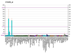
Inducible T-cell costimulator (also called CD278) is an immune checkpoint protein that in humans is encoded by the ICOS (Inducible T-cell COStimulator) gene.[5][6][7][8][9] The protein belongs to the CD28 and CTLA-4 cell-surface receptor family. These are proteins expressed on the surface of immune cells that mediate signalling between them. A surface protein, the ligand, binds specifically to its receptor on another cell, leading to a signalling cascade in that cell.
Function
ICOS is a receptor protein expressed on the surface of activated T cells. Its ligand ICOS-L (previously called B7RP-1) is constitutively expressed on B cells. Stimulation of the ICOS receptor on T cells by ICOS-L on B cells is required for the development of follicular helper T (Tfh) cells.[10]ICOS forms homodimers and plays an important role in cell-cell signaling, immune responses and regulation of cell proliferation.[7]
Knockout phenotype
Compared to wild-type naïve T cells, ICOS-/- T cells activated with plate-bound anti-CD3 have reduced proliferation and IL-2 secretion.[11] The defect in proliferation can be rescued by addition of IL-2 to the culture, suggesting the proliferative defect is due either to ICOS-mediated IL-2 secretion or the activation of similar signaling pathways between ICOS and IL-2. In terms of Th1 and Th2 cytokine secretion, ICOS-/- CD4+ T cell activated in vitro reduced IL-4 secretion, while maintaining similar IFN-g secretion. Similarly, CD4+ T cells purified from ICOS-/- mice immunized with the protein keyhole limpet hemocyanin (KLH) in alum or complete Freund's Adjuvant have attenuated IL-4 secretion, but similar IFN-g and IL-5 secretion when recalled with KLH.
These data are similar to an airway hypersensitivity model showing similar IL-5 secretion, but reduced IL-4 secretion in response to sensitization with Ova protein, indicating a defect in Th2 cytokine secretion, but not a defect in Th1 differentiation as both IL-4 and IL-5 are Th2-associated cytokines. In agreement with reduced Th2 responses, ICOS-/- mice expressed reduced germinal center formation and IgG1 and IgE antibody titers in response to immunization.
Combination therapy
Ipilimumab patients expressed increased ICOS+ T cells in tumor tissues and blood. The increase served as a pharmacodynamic biomarker of anti-CTLA-4 treatment. In wild-type C57BL/6 mice, anti-CTLA-4 treatment resulted in tumor rejection in 80 to 90% of subjects, but in gene-targeted mice that were deficient for either ICOS or its ligand (ICOSLG), the efficacy was less than 50%. An agonistic stimulus for the ICOS pathway during anti-CTLA-4 therapy resulted in an increase in efficacy that was about four to five times as large as that of control treatments. This combination therapy incorporating ICOS costimulation and CTLA-4 blockade effectively remodels tumor-associated macrophages (TAMs) towards an antitumor phenotype, demonstrating promising therapeutic potential in cancer treatment.[12] As of 2015 antibodies for ICOS were not available for clinical testing.[13]
References
Further reading
External links


