West Nile Virus
| West Nile Virus | |
|---|---|
 | |
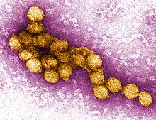
| A micrograph of the West Nile Virus, appearing in yellow | |
| Virus classification | |
| Group: | Group IV ((+)ssRNA) |
| Genus: | |
West Nile Virus (WNV) is a single-stranded RNA virus that causes West Nile fever. It is a member of the family Flaviviridae, specifically from the genus Flavivirus which also contain the Zika virus, dengue virus, and the yellow fever virus. The West Nile virus is primarily transmitted through mosquitoes, mostly by the Culex species. However, ticks have been found to carry the virus. The primary hosts of WNV are birds, remaining within a "bird-mosquito-bird" transmission cycle. [1]
Structure
WNV, like most other flaviviruses, are enveloped viruses, but have icosahedral symmetry.[2] Their envelopes consist of a protein shell and a lipid membrane. The protein shell is made up of two structural proteins: the glycoprotein E and the small membrane protein M.[3] Protein E serve numerous functions, several of which include receptor binding, viral attachment, and entry into the cell through membrane fusion.[4][3]
The flavivirus lipid membrane has been found to contain cholesterol and phosphatidylserine, but other elements of the membrane have yet to have be identified[5][6]. The lipid membrane has many roles in viral infection including acting as signaling molecules and enhancing entrance into the cell.[7] Cholesterol, in particular, plays an integral part to WNV entering a host cell.[8]
Within the viral envelope, the genome is contained within the capsid. The capsid is one of the first proteins created in an infected cell and has been found that that the capsid prevents apoptosis by affecting the Akt pathway.[9] The capsid is a structural protein and its main purpose is to package RNA into the developing viruses.[10]
Genome

WNV is a single stranded, positive sense RNA virus. Its genome is approximately 11,000 nucleotides long and is flanked by a 5' and 3' non-coding stem loop structures.[11] The coding region of the genome encodes for seven nonstructural (NS) proteins , proteins that aren't incorporated into the structure of new viruses, and three structural proteins, proteins that are a part of the virus structure. The WNV genome is first translated into a polyprotein and later cleaved by virus and host proteases into separate proteins (i.e. NS1, C, E).[12]
Structural Proteins
Structural proteins (C, prM/M, E) are capsid, precursor membrane proteins, and envelope proteins respectively.[11] The structural proteins are located at the 5' end of the genome and are cleaved into mature proteins by proteases.
| Structural Protein | Function |
|---|---|
| C | Capsid protein; encloses the RNA genome, packages RNA into immature virions.[10][13] |
| prM/M | Viruses with M protein are infectious: the presence of M protein allows for the activation of proteins involved in viral entry into the cell. prM (precursor membrane) protein is present on immature virions, by further cleavage by furin to M protein, the virions become infectious.[14] |
| E | A glycoprotein that forms the viral envelope, binds to receptors on the host cell surface in order to enter the cell.[15] |
Nonstructural Proteins
Nonstructural proteins consist of NS1, NS2A, NS2B, NS3, NS4A, NS4B, and NS5. These proteins are mainly in viral replication or act as proteases.[13] The nonstructural proteins are located near the 3' end of the genome.
| Nonstructural Protein | Function |
|---|---|
| NS1 | NS1 is a cofactor for viral replication, specifically for regulation of the replication complex.[16] |
| NS2A | NS2A has a variety of functions: it is involved in viral replication, virion assembly, and inducing host cell death.[17] |
| NS2B | A cofactor for NS3 and together forms the NS2B-NS3 protease complex.[13] |
| NS3 | A protease that is responsible for cleaving the polyprotein to produce mature proteins; it can also acts as a helicase.[18] |
| NS4A | NS4A is a cofactor for viral replication, specifically regulates the activity of the NS3 helicase.[19] |
| NS4B | Inhibits interferon signaling.[20] |
| NS5 | The largest and most conserved protein of WNV, NS5 acts as a methyltransferase and a RNA polymerase, though it lacks proofreading properties.[21][13] |
Life Cycle
Once WNV has successfully entered the bloodstream of its host, the envelope protein, E, binds to attachment factors on the host cell, glycosaminoglycans.[15] These attachment factors aid entry into the cell, however, binding to primary receptors are also necessary.[22] Primary receptors include DC-SIGN, DC-SIGN-R, and the integrin αvβ3.[23] By binding to these primary receptors, WNV enters the cell through clathrin-mediated endocytosis.[24] As a result of endocytosis, WNV enters the cell in an endosome.
The acidity of the endosome catalyzes the fusion of the endosomal and viral membranes, allowing the genome to be released into the cytoplasm.[25] Translation of the positive single stranded RNA occurs at the endoplasmic reticulum; the RNA is translated into a polyprotein which is then cleaved by NS2B-N23, viral proteases, to produce mature proteins.[26]
In order to replicate its DNA, NS5, a RNA polymerase, forms a replication complex with other nonstructural proteins to produce an intermediary negative sense single stranded RNA; the negative sense strand serves as a template for synthesis of the final positive sense RNA.[22] Once the positive sense RNA has been synthesized, the capsid protein, C, encloses the RNA strands into immature virions.[23] The rest of the virus is assembled along the endoplasmic reticulum and through the Golgi apparatus, and results in non-infectious immature virions.[26] The E protein is then glycosylated and prM is cleaved by furin, a host cell protease, into the M protein, thereby producing an infectious mature virion.[27][26] The mature viruses are then secreted out of the cell,