

원본 파일 (1,132 × 1,000 픽셀, 파일 크기: 139 KB, MIME 종류: image/jpeg)
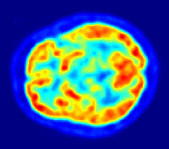
| 설명PET-image.jpg | English: This is a transaxial slice of the brain of a 56 year old patient (male) taken with positron emission tomography (PET). The injected dose have been 282 MBq of 18F-FDG and the image was generated from a 20 minutes measurement with an ECAT Exact HR+ PET Scanner. Red areas show more accumulated tracer substance (18F-FDG) and blue areas are regions where low to no tracer have been accumulated. العربية: صورة مقطعية للدماغ البشري تظهر استهلاك الطاقة. | |||
| 날짜 | ||||
| 출처 | 자작 | |||
| 저자 | Jens Maus (http://jens-maus.de/) | |||
| 저작권 (이 파일을 인용하기) |
|
날짜/시간 링크를 클릭하면 해당 시간의 파일을 볼 수 있습니다.
| 날짜/시간 | 섬네일 | 크기 | 사용자 | 설명 | |
|---|---|---|---|---|---|
| 현재 | 2017년 12월 12일 (화) 11:00 | | 1,132 × 1,000 (139 KB) | SteinsplitterBot | Bot: Image rotated by 270° |
| 2010년 3월 16일 (화) 23:36 |  | 1,002 × 1,132 (134 KB) | Damato | uploaded another PET image with a higher resolution which might be more usable for printing it and which has a better color scale. | |
| 2005년 11월 7일 (월) 18:47 |  | 373 × 405 (48 KB) | Damato | This is an image taken from a typical PET acquisition. It is a tomographic view of a brain examination in transaxial view. Red areas show more accumulated radioactivity and blue areas are partions where low to no activity was accumulated. It should illust |
다음 문서 2개가 이 파일을 사용하고 있습니다:
다음 위키에서 이 파일을 사용하고 있습니다:
이 파일의 더 많은 사용 내역을 봅니다.
이 파일에는 카메라나 스캐너가 파일을 만들거나 디지털화하는 데 사용하기 위해 기록한 부가 정보가 포함되어 있습니다.
프로그램에서 파일을 편집한 경우, 새로 저장한 파일에 일부 부가 정보가 빠질 수 있습니다.
| 방향 | 일반 |
|---|---|
| 수평 해상도 | 600 dpi |
| 수직 해상도 | 600 dpi |
| 사용한 소프트웨어 | Adobe Photoshop CS3 Macintosh |
| 파일이 바뀐 날짜와 시간 | 2010년 3월 16일 (화) 15:34 |
| 색 공간 | sRGB |
| 그림 너비 | 1,002 px |
| 그림 높이 | 1,132 px |
| 날짜와 시간(디지털 데이터) | 2010년 3월 16일 (화) 16:34 |
| 메타데이터가 마지막으로 수정된 날짜 | 2010년 3월 16일 (화) 16:34 |