

원본 파일 (1,420 × 1,091 픽셀, 파일 크기: 259 KB, MIME 종류: image/jpeg)
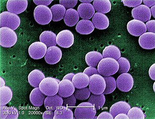
| 설명Staphylococcus aureus VISA 2.jpg | English: Under a very high magnification of 20,000x, this scanning electron micrograph (SEM) shows a strain of Staphylococcus aureus bacteria taken from a vancomycin intermediate resistant culture (VISA). Under SEM, one can not tell the difference between bacteria that are susceptible, or multidrug resistant, but with transmission electron microscopy (TEM), VISA isolates exhibit a thickening in the cell wall that may attribute to their reduced susceptibility to vancomycin . See PHIL 11156 for a black and white version of this image. VISA and VRSA are specific types of antimicrobial-resistant staph bacteria. While most staph bacteria are susceptible to the antimicrobial agent vancomycin some have developed resistance. VISA and VRSA cannot be successfully treated with vancomycin because these organisms are no longer susceptibile to vancomycin. However, to date, all VISA and VRSA isolates have been susceptible to other Food and Drug Administration (FDA) approved drugs. How do VISA and VRSA get their names? Staph bacteria are classified as VISA or VRSA based on laboratory tests. Laboratories perform tests to determine if staph bacteria are resistant to antimicrobial agents that might be used for treatment of infections. For vancomycin and other antimicrobial agents, laboratories determine how much of the agent it requires to inhibit the growth of the organism in a test tube. The result of the test is usually expressed as a minimum inhibitory concentration (MIC) or the minimum amount of antimicrobial agent that inhibits bacterial growth in the test tube. Therefore, staph bacteria are classified as VISA if the MIC for vancomycin is 4-8µg/ml, and classified as VRSA if the vancomycin MIC is >16µg/ml. | ||
| 날짜 | |||
| 출처 |
| ||
| 저자 | Content Providers(s):CDC/ Matthew J. Arduino, DRPH | ||
| 저작권 (이 파일을 인용하기) | PD-USGov-HHS-CDC English: None - This image is in the public domain and thus free of any copyright restrictions. As a matter of courtesy we request that the content provider be credited and notified in any public or private usage of this image. |
| Public domainPublic domainfalsefalse |
This image is a work of the Centers for Disease Control and Prevention, part of the United States Department of Health and Human Services, taken or made as part of an employee's official duties. As a work of the U.S. federal government, the image is in the public domain. eesti ∙ Deutsch ∙ čeština ∙ español ∙ português ∙ English ∙ français ∙ Nederlands ∙ polski ∙ slovenščina ∙ suomi ∙ македонски ∙ українська ∙ 日本語 ∙ 中文(简体) ∙ 中文(繁體) ∙ العربية ∙ +/− |
날짜/시간 링크를 클릭하면 해당 시간의 파일을 볼 수 있습니다.
| 날짜/시간 | 섬네일 | 크기 | 사용자 | 설명 | |
|---|---|---|---|---|---|
| 현재 | 2009년 8월 4일 (화) 11:24 | | 1,420 × 1,091 (259 KB) | Raeky | {{Information |Description={{en|1='''Under a very high magnification of 20,000x, this scanning electron micrograph (SEM) shows a strain of Staphylococcus aureus bacteria taken from a vancomycin intermediate resistant culture (VISA).'''<p> Under SEM, one |
다음 문서 1개가 이 파일을 사용하고 있습니다:
다음 위키에서 이 파일을 사용하고 있습니다:
이 파일의 더 많은 사용 내역을 봅니다.
이 파일에는 카메라나 스캐너가 파일을 만들거나 디지털화하는 데 사용하기 위해 기록한 부가 정보가 포함되어 있습니다.
프로그램에서 파일을 편집한 경우, 새로 저장한 파일에 일부 부가 정보가 빠질 수 있습니다.
| _error | 0 |
|---|